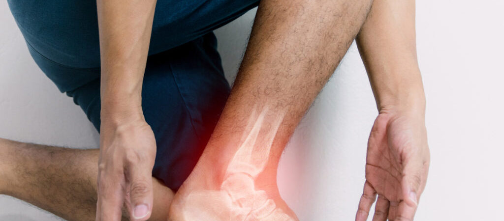
Peroneal Tendonitis condition develops when peroneal tendons located in the legs get inflamed due to friction and thicken in size for handling the pressure. Highly active people and athletes visit Dr. Sima at the Podiatrist Irvine Clinic in Orange County for diagnosis and treatment. The symptoms are similar to Plantar Fasciitis but the inflamed tendons are located on the sides of your foot instead of on the bottom.
Anatomy
Tendon is a group of tissue that connects the muscles and bones. There are two tendons called Peroneal Brevis and Peroneal Longus tendons that run parallel on the exterior side of the foot. The tendons are just behind the Fibula bone on the outside of your ankle called the Lateral Malleus. One tendon is attached to the base of the little toe on the outside of your foot. The other tendon is attached to the arch from underneath the foot.
Ankle gets the support and stability from the peroneal tendons to handle the weight and protect from sprains. It even helps you to turn your foot out and keep the arch steady while walking.
Possible causes of peroneal tendonitis
Peroneal Tendonitis is the thickening, swelling, and enlargement of these tendons. The causes of this condition include –
- Overuse of ankle motion
- Sudden increase in weight-bearing activities
- Poor training techniques
- Improper footwear
- Frequently spraining the ankle
- Poor foot biomechanics
- High foot arches
- Imbalanced muscles in lower limbs
- Lower limb joints and muscles not working in sync
Symptoms of peroneal tendonitis
- Ankle pain worsens during physical activity
- Pain when ankle rotates
- Ankle area swelling
- Ankle instability while bearing weight
- The ankle area feels warm
- Pain & stiffness when foot is rolled outward
How does the podiatrist treat Peroneal Tendonitis?
Irvine Podiatrist discusses the medical history, which will often be about increased activity, overuse, or associated causes of the condition. It is crucial to diagnose if the pain is because of peroneal tendons and not associated with fibula as this deciphers a different issue. There are two stages of peroneal tendon pain.
- The acute stage means you have not experienced pain before. The peroneal tendon pain is there for less than 5 to 6 weeks. It can be due to new activities like running uphill or hiking or having suffered an acute ankle sprain.
- In the subacute stage, the pain has been persistent from 3 to 6 months. The patients have been on some kind of conservative therapy but there is no significant improvement. In this phase, intratendinous pathology starts to occur. As months pass the tendon suffers from chronic scarring and different stages of cavus foot deformity. Severe cavus foot deformity means the antagonistic muscles have crushed the peroneals.
A physical exam includes a comprehensive assessment of –
- Vascular status
- Skin envelope
- Neuromuscular systems
If the podiatrist doubts progressive neuromuscular disease a neurological workup is recommended. During the musculoskeletal examination, the peroneal tendons are palpated to find the pain severity location edema/inflammation and palpable thickening of the tendon itself.
During active eversion, tendon subluxation [dislocation, joint stability, ankle motion, and even foot structure is evaluated. If there is varus deformity then the Coleman block test is done to determine deformity flexibility and reducibility.
Conservative treatment
For acute peroneal tendonitis
If there is an acute subluxation or peroneal tear the patient is recommended the R.I.C.E. therapy [rest, ice, compression, and elevation]. The Orange County podiatrist can also recommend immobilization crucial for ankle support to protect the peroneal tendon from overload until it heals. Immobilization involves using snug-fitting braces, ankle taping, or wearing ankle boots.
Physical therapy is also crucial to increase strength and decrease inflammation. Patients with ankle instability can benefit from proprioception training. If patients have the timely treatment they will see improvement within a month.
Unfortunately, if there is no or little improvement using the above conservative treatment after a couple of months, the podiatrist recommends an MRI. It helps them evaluate the tendon and its ligament makeup.
If they detect longitudinal tearing or thickening of one/both tendon/s a PRP [Platelet-rich plasma] or amniotic therapy is performed. The injections are administered followed by boot immobilization for two weeks. If the pain persists after four weeks there is a follow-up of PRP or amniotic injection.
For a subtle to severe rigid cavus foot use orthotic support as recommended –
- For mild and flexible cavus foot choose an orthotic with first ray cutout and lateral foot.
- For rigid deformity, AFO or ankle-foot orthosis is great because it prevents painful motions as well as protects from reverse injuries.
- For pes planus foot, n orthotic device helps to prevent the peroneal tendons spasm and overuse pathology occurrence.
- Ankle bracing and physical therapy helps to control ankle instability
If the conservative treatment fails then consider surgical repair.
Surgical treatment
Chronic peroneal tendinopathy
Patients in the chronic peroneal tendinopathy stage will hardly improve with conservative treatment but will need correction of deforming forces. If there is a tear or split that runs along the tendons length surgery is considered to repair the tendons. A deep groove is made in the back of the fibula bone, which gives tendons space to move smoothly. If the tendon is diseased, the surgeon will have to resect and connect both bravis and longus together. Some surgery is complicated depending on the pathology level of the tendons.
The majority of patients recover with conservative therapy, activity changes, and footwear modification. The root cause of Peroneal tendon pain is not just a ligamentous injury, so it is essential to visit a local podiatrist for a perfect diagnosis and prevent grave foot deformities.


